Imaging neuroscience
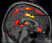
This research area is concerned with the application of imaging and computational methods to understand brain function in health and disease.
We are at the forefront of developing methods for use in brain research, as well as working closely with collaborators to use these methods to understand and interpret brain function in diverse areas. These include:
- Semantic and vascular dementia and normal ageing
- Anti-social personality disorder
- Depression and schizophrenia
- Gut-brain interactions
- Cortical plasticity
- Stroke
The aim is to develop and evaluate novel biomarkers that allow tissue or lesion status to be defined non-invasively, thereby allowing disease states to be characterised and treatment response to be quantified.
As such, the activities tie in closely with the core methodological biomarker development research undertaken in Imaging Sciences, and can be considered as both a translational output of that group, but also an intelectual and scientific driver of its activities.
Summary of current active research
Professor Steve Williams
Development and application of MR spectroscopy and functional imaging methods to understand neurological conditions and the effects of pharmaceutical intervention.
Collaborators include:
-
Professor Bill Deakin and colleagues in the Neuroscience and Psychiatry Unit
Investigating the biological basis of psychiatric diseases, with a particular interest in combining drug-challenge with functional MRI (pMRI), and investigating connectivity using resting-state blood oxygenation (BOLD) measurements -
Dr Shaheen Hamdy
GI Sciences using MRS to research the role of neurotransmitters in experimentally induced cortical plasticity -
Professor Simon Luckman, Faculty of Life Sciences
Investigating appetite regulation using pMRI in animal models -
Professor Anthony Jones, Human Pain Research Group
Functional MRI of psychological modeulations of the response to pain
Professor Tim Cootes
The application of statistical models of shape and appearance to the analysis of images of the brain, in particular for detecting and quantifying shape changes and for accurate location of structures within the brain.
Developing automated methods for constructing statistical atlases of brain shape and appearance, which represent the variation of shape across populations.
Dr Neil Thacker
Since 2000, Dr Thacker has led a group developing software for fully quantitative, statistically based, methods for automated anayisis of medical images.
Techniques developed so far include:
- quantitation of tissue from MR images of the head
- volumetric data alignment
- RF field inhomogeneity correction
- MR based fMRI analysis
- MR spectroscopic data analysis
- analysis of cerebro spinal fluid pulsitility and microvascular perfusion.
A full description of this work and software is available from the TINA website.
These techniques have been developed for, and applied in, a number of scientific studies which investigated degeneration of the brain tissues and associated physiology in disease and normal ageing. Comparisons have also been performed between these results and equivalent techniques available in the literature. In addition Dr Thacker has co-ordinated fMRI studies to investigate brain function in high-level vision for comparison with computational models.
Professor Geoff Parker
Professor Parker leads a group researching cerebral network structure and function, with an emphasis on tractography using diffusion weighted MRI.
This group is involved in the use of tractography to understand structure-function relationships in the healthy human brain, and how these relationships are altered in disease states.
Current topics of research include:
- improved acquisition strategies for tractography and fMRI
- novel quantitative tractography techniques
- development of quantitative perfusion and structural imaging methods.
Current clinical applications, involving collaborators within Manchester and beyond, include dementias, stroke, epilepsy, cancer, and schizophrenia.

