Imaging biomarkers and signal-based biomarkers
This research area is concerned with the broad development of image and signal-based biomarkers of tissue status in health and disease.
The aim is to develop and evaluate novel biomarkers that allow tissue or lesion status to be defined non-invasively, thereby allowing disease states to be characterised and treatment response to be quantified.
These activities form the core methodological biomarker development research undertaken in ISBE and support our programmes of work in multiple disease areas through a programme of translation and application of new methodologies and technologies.
Summary of current active research
Professor Geoff Parker
Development and application of imaging biomarkers in cancer, neuroscience, and inflammatory conditions.
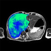
Professor Parker heads the Quantitative Biomedical Imaging Lab in the Imaging Sciences Research Group, which is dedicated primarily to the development and application of imaging biomarkers including blood flow, capillary permeability, and tissue oxygenation measurements for therapeutic trials of anti-cancer agents.
Professor Parker also leads a group dedicated to the development of biomarkers of cerebral network structure and function, with a particular emphasis on tractography using diffusion weighted MRI. This group is involved in the use of tractography to understand structure-function relationships in the healthy human brain and how these relationships are altered in disease states.
Professor Alan Jackson
Development and application of imaging biomarkers of human micro- and macro-vasculature and of tissue atrophy in cancer and vascular diseases.
Professor Tim Cootes
Development and application of statistical models of shape and image texture for understanding structural and functional variation in health and diseased states.
Professor Cootes has expertise in developing statistical models of the variation in shape and image appearance across populations. Such models can compactly represent both the normal variation and changes due to disease.
Dr Jim Graham
Professor Chris Taylor
Dr Neil Thacker
Professor Steve Williams
Development and application of image analysis methods for understanding structural and functional variation in health and disease.
Development and application of image modelling methods for understanding structural and functional variation in health and disease.
Development and application of image analysis methods to quantify cerebral tissue atrophy and blood flow.
Development and application of MR spectroscopy and functional imaging methods to understand neurological conditions and the effects of pharmaceutical intervention.